Chimeric antigen receptor (CAR) T-cell therapy is effective in relapsed/refractory large B-cell lymphoma and results in a unique toxicity profile, namely cytokine release syndrome (CRS) and immune effector cell-associated neurotoxicity syndrome. The hyper-inflammatory state associated with these toxicities has been suggested to increase the risk of thrombosis.
We conducted a retrospective analysis of patients treated with axicabtagene ciloleucel (axi-cel) to assess the rate of thrombosis with axi-cel therapy from the time of CAR T-cell infusion until the end of hospitalization, when performed in the inpatient setting, or up to day +30 when performed in the outpatient setting. Ninety-two (95%) of 97 patients were hospitalized during axi-cel therapy and 85 (88%) developed CRS. Fifty-five patients (57%) received concurrent anticoagulation (53 as prophylaxis). Patients with prior VTE did not have progression https://joplink.net/antigens/ or evidence of new VTE. Only 2 (2.1%) patients developed VTE. These results demonstrate a low-risk for thrombosis in axi-cel recipients.
The Protective Action of Piperlongumine Against Mycobacterial Pulmonary Tuberculosis in Its Mitigation of Inflammation and Macrophage Infiltration in Male BALB/c Mice
Introduction: Piperlongumine (PL) is a bioactive alkaloid and medicinal compound of piperamide isolated from the long pepper (Piper longum Linn). It has demonstrated bactericidal action against Mycobacterium tuberculosis (MTB), the cause of pulmonary tuberculosis; nevertheless, immunomodulatory activity had not been identified for it in MTB-triggered granulomatous inflammation. This study investigated if piperlongumine could inhibit such inflammation.
Material and methods: Mycobacterium tuberculosis strain H37Rv was subjected to a broth microdilution assay. Piperlongumine at 5, 15, and 25 μg/mL, 0.2% dimethyl sulphoxide as control or 4 μM of dexamethasone were tested in vitro on MH-S murine alveolar macrophages. BALB/c mice were orally administered PL at 50, 100 and 150 mg/kg b.w. after trehalose-6,6-dimycolate (TDM) stimulation.
Chemokine and cytokine concentrations were determined in lung supernatants. Flow cytometry and Western blot analysis were performed to determine phosphorylated spleen tyrosine kinase (Syk), c-Jun N-terminal kinase (JNK) and extracellular signal-regulated kinase (ERK) pathways.
Results: Piperlongumine inhibited inflammatory mediators and adherence of lymphocyte function-associated antigen 1 to MH-S cells following TDM activation. It also improved macrophage clearance of MTB. In TDM-stimulated MH-S cells, PL significantly influenced the macrophage inducible Ca2+-dependent lectin receptor (Mincle)-Syk-ERK signalling pathway. Oral dosing of PL effectively suppressed the development of pulmonary granulomas and inflammatory reactions in the TDM-elicited mouse granuloma model.
Prevalence of Toxoplasma Gondii in Retail Fresh Meat Products from Free-range Chickens in Spain
Toxoplasma gondii is one of the most prevalent zoonotic protozoan parasites worldwide and affects the vast majority of warm-blooded animal species, including humans. Postnatal infection in humans occurs through the ingestion of sporulated T. gondii oocysts or via the oral intake of parasite tissue cysts during the consumption of raw or undercooked meat. In this regard, given their high exposure to oocysts, chickens (Gallus domesticus) raised on the ground constitute a potential source of T. gondii.
Material and methods: For the first time in Spain, a survey was undertaken in commercial retail free-range poultry. A total of 50 thighs from different animals were analysed. The samples were homogenised and an acid pepsin digestion procedure was applied prior to molecular analysis. Toxoplasma gondii DNA was isolated from meat by qPCR. Two sets of primers were used for DNA amplification targeting the specific sequence of a 529 bp repeat element and another set of primers was utilised for the surface antigen protein-1 gene.
Interrelationships between amphiregulin, kisspeptin, FSH and FSH receptor in promotion of human ovarian cell functions
The aim of this study was to investigate: (1) the ability of granulosa cells to produce amphiregulin (AREG), kisspeptin (KISS) and FSH receptor (FSHR); (2) the role of AREG and KISS in the control of ovarian functions; (3) the effect of FSH and KISS on AREG; and (4) the ability of KISS to affect FSHR and to modify FSH action on AREG output by human ovarian granulosa cells. We examined: (1) time-dependent accumulation of AREG; (2) effects of AREG (0, 1, 10, 100ng/mL) and KISS (0, 1, 10, 100ng/mL) on granulosa cell functions; and (3) the effects of KISS (0, 1, 10, 100ng/mL), FSH (0, 1, 10, 100ng/mL), and their combinations on AREG release.
Viability, markers of proliferation [accumulation ofproliferating cell nuclear antigen (PCNA) cyclin B1 and sodium 3′-[1-(phenylaminocarbonyl)-3,4-tetrazolium]-bis(4-methoxy6-nitro)benzene sulfonic acid hydrate (XTT formazan)] and apoptosis (accumulation of bax, caspase 3 and terminal deoxynucleotidyl transferase dUTP nick-end labelling), accumulation of KISS, FSHR and steroid hormones, and AREG release were analysed by Trypan blue exclusion test, quantitative immunocytochemistry, XTT, terminal deoxynucleotidyl transferase dUTP nick-end labelling assays and enzyme-linked immunosorbent assay.
AREG promoted cell viability, proliferation and steroid hormone output, and inhibited apoptosis. KISS (1 and 10ng/mL) stimulated viability, proliferation, steroid hormone release and occurrence of FSHR and suppressed apoptosis and AREG output; KISS (100ng/mL) had the opposite effect. FSH stimulated AREG release, whilst addition of KISS reversed this FSH effect. FSH mimicked and promoted the inhibitory effect of KISS on AREG release. These results suggest an intra-ovarian production and a functional interrelationship between AREG, KISS, FSH and FSHR in direct regulation of basic ovarian cell functions.
Novel PE and APC tandems: Additional near-infrared fluorochromes for use in spectral flow cytometry
Recent advances in flow cytometry instrumentation and fluorochrome chemistries have greatly increased fluorescent conjugated antibody combinations that can be used reliably and easily in routine experiments. The Cytek Aurora flow cytometer was first released with three excitation lasers (405, 488, and 640 nm) and incorporated the latest Avalanche Photodiode (APD) technology, demonstrating significant improvement in sensitivity for fluorescent emission signals longer than 800 nm.
However, there are limited commercially available fluorochromes capable of excitation with peak emission signals beyond 800 nm. To address this gap, we engineered six new fluorochromes: PE-750, PE-800, PE-830 for the 488 nm laser and APC-750, APC-800, APC-830 for the 640 nm laser.
Utilizing the principal of fluorescence resonance energy transfer (FRET), these novel structures were created by covalently linking a protein donor dye with an organic small molecule acceptor dye. Additionally, each of these fluorochrome conjugates were shown to be compatible with fixation/permeabilization buffer reagents, and demonstrated acceptable brightness and stability when conjugated to antigen-specific monoclonal antibodies. These six novel fluorochrome reagents can increase the numbers of fluorochromes that can be used on a spectral flow cytometer.
Paraneoplastic and Other Autoimmune Encephalitides: Antineuronal Antibodies, T Lymphocytes, and Questions of Pathogenesis
Autoimmune and paraneoplastic encephalitides represent an increasingly recognized cause of devastating human illness as well as an emerging area of neurological injury associated with immune checkpoint inhibitors. Two groups of antibodies have been detected in affected patients. Antibodies in the first group are directed against neuronal cell surface membrane proteins and are exemplified by antibodies directed against the N-methyl-D-aspartate receptor (anti-NMDAR), found in patients with autoimmune encephalitis, and antibodies directed against the leucine-rich glioma-inactivated 1 protein (anti-LGI1), associated with faciobrachial dystonic seizures and limbic encephalitis. Antibodies in this group produce non-lethal neuronal dysfunction, and their associated conditions often respond to treatment.
Antibodies in the second group, as exemplified by anti-Yo antibody, found in patients with rapidly progressive cerebellar syndrome, and anti-Hu antibody, associated with encephalomyelitis, react with intracellular neuronal antigens.
These antibodies are characteristically found in patients with underlying malignancy, and neurological impairment is the result of neuronal death. Within the last few years, major advances have been made in understanding the pathogenesis of neurological disorders associated with antibodies against neuronal cell surface antigens.
In contrast, the events that lead to neuronal death in conditions associated with antibodies directed against intracellular antigens, such as anti-Yo and anti-Hu, remain poorly understood, and the respective roles of antibodies and T lymphocytes in causing neuronal injury have not been defined in an animal model.
In this review, we discuss current knowledge of these two groups of antibodies in terms of their discovery, how they arise, the interaction of both types of antibodies with their molecular targets, and the attempts that have been made to reproduce human neuronal injury in tissue culture models and experimental animals.
We then discuss the emerging area of autoimmune neuronal injury associated with immune checkpoint inhibitors and the implications of current research for the treatment of affected patients.
The Rapid Response COVID-19 Antigen Rapid Test Pen (Saliva) is an in vitro immunoassay. The assay is for the direct and qualitative detection of SARS-CoV-2 viral nucleoprotein antigens from saliva samples through visual interpretation of colour development. This test is intended for professional use only.
Key Benefits:
• First and the only no-spit test dedicated to COVID-19 – Its sponge-like pen tip will collect a sufficient volume of specimen in 2 mins without the need to spit and avoiding uncomfortable nasal/throat swabbing.
• ALL IN ONE – No need to waste time setting up a workstation and handle numerous assay components. All you need is one pen that can be used comfortably in any location.
Test Principle
Anti-SARS-CoV-2 antibodies are immobilized on the test region of the nitrocellulose membrane. Anti-SARS-CoV-2 antibodies conjugated to coloured particles are immobilized on the conjugated pad. A sample is added to the extraction buffer which is optimized to release the SARS-CoV-2 antigens from specimen. During testing, target antigens, if present in the saliva samples, will be released into the extraction buffer individually packed in the kit. Consequently, the extracted antigens will bind to anti-SARS-CoV-2 antibodies conjugated to coloured particles
As the specimen migrates along the strip by capillary action and interacts with reagents on the membrane, the complex will be captured by the anti-SARS-CoV-2 antibodies at the test region. Excess coloured particles are captured at the internal control zone. The presence of a coloured band in the test region indicates a positive result for the SARS-CoV-2 viral antigens, while its absence indicates a negative result. A coloured band at the control region serves as a procedural control, indicating that the proper volume of specimen has been added and membrane wicking is working.
ï¿ INVBIO saliva alcohol screening test kit detects alcohol presence in saliva. Therefore, directly dipping the strip into alcohol alone will not provide you with an accurate reading.
ï¿ It is very important that the test be read at exactly two minutes, The result you read 2 minutes after saturation with saliva is the accurate test result. ï¿Nothing should be placed into the mouth of the subject for at least 10 minutes prior to saliva collection. This includes food, drink, tobacco products or other materials.
SPECIMEN COLLECTION AND PREPARATION
ï¿Nothing should be placed into the mouth of the subject for at least 10 minutes prior to saliva collection. This includes food, drink, tobacco products or other materials.
ï¿ Saliva specimen can be collected in a sputum cup or a clean container, or directly applied to the reaction pad of the test strip.
PROCEDURE:
Open the foil package and remove the test strip.
Saturate the reactive pad by dipping the reaction pad into the saliva specimen collected in a cup, or saturate the reactive pad on the end of stick with saliva in mouth for 10 seconds, shake off the excess saliva.
Immediately start timer and at exactly 2 minutes, compare the reactive pad with the provided colored chart.
Results after more than 2 minutes may be not accurate.
INTERPRETATION OF RESULTS:
Negative: Almost no color change by comparing with the background. The negative result indicates that the BAC is less than 0.02%.
Positive: A distinct color developed all over the pad. The positive result indicates that the BAC is 0.02% or higher.
Invalid: The test should be considered invalid If only the edge of the reactive pad turned color that might be ascribed to insufficient sampling.
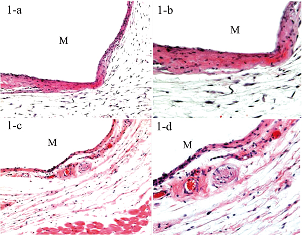
We aimed to guage the subcutaneous tissue response to a newly developed adhesive silicone denture relining materials, SG, (Neo Dental Chemical Products Co., Ltd. Tokyo, Japan). We embedded the experimental materials SG and one other present management materials, Roeko Seal (RS), within the dorsal space of 22 male ddY mice.
One week and 12 weeks after the embedding, the tissues surrounding the embedded supplies had been eliminated and a histopathological examination was carried out. The outcomes reveal that the essential histopathological elements are the formation of granulation tissue and the change of the tissue to fibrous capsule over time.
The outcomes means that the newly-developed SG is secure as in contrast with the management RS, whose composition is comparable.
Histopathological examination of newly-developed adhesive silicone denture relining materials.
Collaborative research on fifteen compounds within the rat-liver Comet assay built-in into 2- and 4-week repeat-dose research.
A collaborative trial was carried out to guage the likelihood of integrating the rat-liver Comet assay into repeat-dose toxicity research. Fourteen laboratories from Europe, Japan and the USA examined fifteen chemical compounds.
Two chemical compounds had been beforehand proven to induce micronuclei in an acute protocol, however had been discovered unfavorable in a 4-week Micronucleus (MN) Assay (benzo[a]pyrene and 1,2-dimethylhydrazine; Hamada et al., 2001); 4 genotoxic rat-liver carcinogens that had been unfavorable within the MN assay in bone marrow or blood (2,6-dinitrotoluene, dimethylnitrosamine, 1,2-dibromomethane, and 2-amino-3-methylimidazo[4,5-f]quinoline); three compounds used within the ongoing JaCVAM (Japanese Center for the Validation of Alternative Methods) validation research of the acute liver Comet assay (2,4-diaminotoluene, 2,6-diaminotoluene and acrylamide); three pharmaceutical-like compounds (chlordiazepoxide, pyrimethamine and gemifloxacin), and three non-genotoxic rodent liver carcinogens (methapyrilene, clofibrate and phenobarbital). Male rats obtained oral administrations of the take a look at compounds, each day for 2 or 4 weeks.
Description: A polyclonal antibody against EDEM2. Recognizes EDEM2 from Human. This antibody is Unconjugated. Tested in the following application: ELISA, IHC; Recommended dilution: IHC:1:20-1:200
Description: A polyclonal antibody against EDEM2. Recognizes EDEM2 from Human. This antibody is Unconjugated. Tested in the following application: IHC, ELISA;IHC:1/100-1/300.ELISA:1/10000
Description: A polyclonal antibody for detection of EDEM2 from Human. This EDEM2 antibody is for IHC-P, ELISA. It is affinity-purified from rabbit antiserum by affinity-chromatography using epitope-specific immunogenand is unconjugated. The antibody is produced in rabbit by using as an immunogen synthesized peptide derived from the C-terminal region of human EDEM2
Description: A polyclonal antibody for detection of EDEM2 from Human. This EDEM2 antibody is for IHC-P, ELISA. It is affinity-purified from rabbit antiserum by affinity-chromatography using epitope-specific immunogenand is unconjugated. The antibody is produced in rabbit by using as an immunogen synthesized peptide derived from the C-terminal region of human EDEM2
Description: A polyclonal antibody for detection of EDEM2 from Human. This EDEM2 antibody is for IHC-P, ELISA. It is affinity-purified from rabbit antiserum by affinity-chromatography using epitope-specific immunogenand is unconjugated. The antibody is produced in rabbit by using as an immunogen synthesized peptide derived from the C-terminal region of human EDEM2
Description: A polyclonal antibody raised in Rabbit that recognizes and binds to Human EDEM2 . This antibody is tested and proven to work in the following applications:
Description: A polyclonal antibody against EDEM2. Recognizes EDEM2 from Human. This antibody is HRP conjugated. Tested in the following application: ELISA
Description: A polyclonal antibody against EDEM2. Recognizes EDEM2 from Human. This antibody is FITC conjugated. Tested in the following application: ELISA
Description: A polyclonal antibody against EDEM2. Recognizes EDEM2 from Human. This antibody is Biotin conjugated. Tested in the following application: ELISA
Description: Human EDEM2 knockdown cell line is engineered by our optimized transduction of the specific shRNA with lentivirus. Knockdown levels are determined via qRT-PCR. Gentaur offers generation of stable knockdown (RNAi) cell lines expressing shRNAs targeting genes of your interest.
Description: A Monoclonal antibody against Human EDEM2 (clone 2E4). The antibodies are raised in Mouse and are from clone 2E4. This antibody is applicable in WB and IHC-P, E
Description: Gentaur can provide custom lentiviral constructs expressing any genes of interest as long as it is less than about 3 kb. Lentiviral technology enables us to efficiently generate stable expression lines which are then selected for moderate or high expressers, depending on the experimental requirements. If you are interested in specific lentiviral DNA constructs or have further questions, please contact us to discuss the details. EDEM2 ER degradation enhancer, mannosidase alpha-like 2 [ Homo sapiens ] http://www.ncbi.nlm.nih.gov/gene/55741
The prime dose was meant to be the very best dose producing scientific indicators or histopathological results with out inflicting mortality, i.e. the 28-day most tolerated dose. The liver Comet assay was carried out in accordance with revealed suggestions and following the protocol for the continuing JaCVAM validation trial. Laboratories supplied liver Comet assay knowledge obtained on the finish of the long-term (2- or 4-week) research along with an analysis of liver histology.
Most of the take a look at compounds had been additionally investigated within the liver Comet assay after short-term (1-Three each day) administration to match the sensitivity of the 2 research designs. MN analyses had been carried out in bone marrow or peripheral blood for many of the compounds to find out whether or not the liver Comet assay might complement the MN assay for the detection of genotoxins after long-term therapy.
Most of the liver genotoxins had been optimistic and the three non-genotoxic carcinogens gave unfavorable end result within the liver Comet assay after long-term administration. There was a excessive concordance between short- and long-term Comet assay outcomes. Most compounds when examined as much as the utmost tolerated dose had been accurately detected in each short- and long-term research.
Discrepant outcomes had been obtained with 2,6 diaminotoluene (unfavorable within the short-term, however optimistic within the long-term research), phenobarbital (optimistic within the short-term, however unfavorable within the long-term research) and gemifloxacin (optimistic within the short-term, however unfavorable within the long-term research).
The total outcomes point out that the liver Comet assay will be built-in inside repeat-dose toxicity research and effectively enhances the MN assay in detecting genotoxins. Practical elements of integrating genotoxicity endpoints into repeat-dose research had been evaluated, e.g. by investigating the impact of blood sampling, as usually carried out throughout toxicity research, on the Comet and MN assays.
The bleeding protocols used right here didn’t have an effect on the conclusions of the Comet assay or of the MN assays in blood and bone marrow. Although bleeding typically elevated reticulocyte frequencies, the sensitivity of the response within the MN assay was not altered.